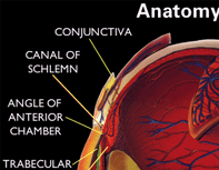
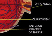
Cornea:
The cornea is a dime-sized clear tissue covering the front of the
eye
which acts like a camera's
aperture & referred
to as the front window of the eye'. It provides most of the focusing
power when light enters your eye. Light rays pass through the cornea
and then through the lens. The lens forms an image on the retina
in the back of the eye where the optic nerve is located. Sight is
controlled by the optic nerve, the only nerve of vision. This nerve
activates the retina to pick up the image in view. The cornea is
transparent structure which is
composed of 5 layers of tissue. The outer layer (the epithelium)
is the eye's protective layer. This layer is made up of highly regenerative
cells that can grow back within 3 days, and therefore, allow for
fast healing of superficial injuries. Most of the inner layers provide
strength to the eye. The corneal grafting,
laser vision or R.K correction procedure is performed on this part
of the eye.
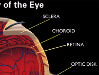
Sclera:
This is the 'white part' that we see
in people's eye. The sclera's purpose is to provide structure, strength
and protection to the eye.
Iris:
The colored part of the eye mostly brown, black & blue. The iris
helps in regulating the amount of light that enters the eye with
contraction or expansion of the muscles of the iris.
Pupil:
Acts like a shutter of camera, situated
in the middle of the iris. The pupil determines how much light is
let into the eye. It changes sizes to accommodate for the amount
of light that is available.
When you are in a bright environment, the pupil becomes smaller
to allow less light through. When it is dark, the pupil expands
to allow more light to reach the back of the eye.
Lens:
The transparent structure located
behind the pupil which helps in focusing light rays over
the retina.
As people reach their 60s or 70s, the lens sometimes becomes cloudy
and hard (cataract formation), preventing
light from entering the eye
Retina:
The nerve layer that lines
the back of the eye. The retina senses light and creates impulses
that are sent through the optic nerve to the brain, acting as film
to record the light (the photo itself).
Macula:
A small area in the retina that contains special light-sensitive
cells. The macula allows us to see fine details clearly.
Optic Nerve:
The nerve that connects the eye to the brain. The optic nerve carries
the impulses formed by the retina to the brain, which interprets
them as images.
Vitreous:
The clear, jelly-like substance that fills the middle of the eye.
Other eye structures:
Support the main activity of sight: Some carry fluids (such as tears
and blood) to lubricate or nourish the eye.
Others are muscles:
That allow the eye movements. Some protect the eye from injury (such
as the eyelids and the epithelium of the cornea). And some are messengers,
sending sensory information to the brain (such as the pain-sensing
nerves in the cornea and the optic nerve behind the retina).