|
Glaucoma is a "silent killer" like Diabetic Retinopathy.
It
is commonly called as Kala Motia in vernacular.
In most cases it begins un-noticeably and damages the eyes
without any sign or symptom till it is very late. This is the
reason that awareness about glaucoma and its treatment is
important to prevent this blinding disease.
What is Glaucoma?
Glaucoma has been rightly called the “The silent stealer of eyesight”.
Glaucoma is a group of disorders in which [I.O.P] intra-ocular
pressure (I.O.P maintains the shape of the eye ) is raised above
the normal value (11 - 21 mm Hg) in the affected eye , resulting
in a damage to the optic nerve head & irreversible visual field
defects.
The loss of vision is so gradual from along the periphery of the
eye that the patient is largely unaware of the loss. What is most
tragic is that vision loss due to glaucoma is irreversible. Medication
and surgery can at the best preserve the remaining eyesight of
the patient.
Glaucoma is a result of build up of fluid within the eye and the
resultant increase in pressure. This pressure falls on the sensitive
optic nerve resulting in irreversible damage.
What are the risk factors for Glaucoma?
Following are the
risk factors for Glaucoma:
1. Family history
of glaucoma especially in parents and siblings (risk of 10% in
siblings).
2. Diabetes mellitus
3. Thyroid Diseases: Thyroiditis
4. Refractive error
:mostly seen in high Myopes
5. Cigarette smoking
6. Injudicious use
of steroids (eye drops)
7. Age: Mostly glaucoma
affect people in the fifth & seventh decade of their life but
it can occur at any age.
Can glaucoma can occur in children?
Glaucoma
can occur even in young children and infants (Developmental/Congenital
Glaucoma). Occurring before the age of 3 years it is called Congenital
Glaucoma.
Between the age of 3 and 30-35 years it is called Juvenile Glaucoma.
Types of Glaucoma- There are two main
types of glaucoma:
Open Angle Glaucoma & Angle
Closure Glaucoma
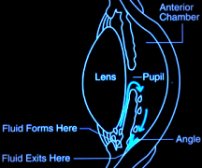
Normally, the fluid
of the eye (aqueous) circulates through anterior chamber and passes
through the angle exits from the eye into the Canal of Schlemn
but in open angle glaucoma the passage to the canal of Schlemn
offer resistance to the flow of aqueous.
 Chronic
Open Angle glaucoma Chronic
Open Angle glaucoma
As there are hardly any symptoms, the patient is largely unaware
that there is a progressive loss of vision. The only way of diagnosis
is through periodic eye examinations.
 Congenital
Glaucoma Congenital
Glaucoma
This is visible literally from birth. Since the eye of an infant
is more elastic than that of an adult, the increase of pressure
leads to the eye bulging out.
 Acute Angle Closure Glaucoma
This is a sudden onset. Blurred vision, severe pain, rainbow
haloes around light, nausea and vomiting are the immediate symptoms.
Unless medical attention is provided immediately, blindness can
result in a day or two.
Anybody over the age of 40 years is susceptible to Glaucoma. That’s
why it is advised that people above 40 should go in for an ophthalmologic
examination every 6 months.

In angle closure glaucoma
the angle of the chamber is narrow or gets closed preventing the
drainage of aqueous from the eye. Both types lead to increase
in intra-ocular pressure.
What are the signs & symptoms?
Primary open angle glaucoma usually insidious & asymptomatic
(does not give rise to any symptoms) in early stages.
In late stages patients may feel pain in eyes (eye-ache).
Some individuals may
notice field defects (inability to see certain areas of the field
of vision).
Usually
this type of glaucoma is diagnosed on examination by an eye specialist.
Patient with very high I.O.P may complain of occasional colored
rings (haloes) around lights due to transient corneal epithelial
oedema.
Patient can develop
delayed dark adaptation.
Reading & close work often present increasing difficulties owing
to accommodative failure due to constant pressure on the ciliary
muscle & it is nerve supply. Therefore patient usually complain
of frequent changes in presbyopic glasses.
How glaucoma can lead to blindness?
Increase in pressure
in the eye leads to resistance to flow of blood into the eye leading
to damage to the
optic nerve head which carries the images to the brain. First
it leads to some area of loss of visual field (the extent of surrounding
visible to any one eye). This field loss progresses gradually
till the eye is completely blind. Early field loss can be detected
by an eye specialist with a test called Visual Field Charting.
How to diagnose
Glaucoma ?
Glaucoma can be diagnosed
by following tests:
Intra-ocular Pressure
[I.O.P] or Eye Pressure: Measured by an instrument
called Applanation Tonometer (Normal I.O.P ranges between 11 and
21 mm Hg). It may be mildly raised in open angle glaucoma and
markedly raised in angle closure glaucoma.
In between the attacks of angle closure glaucoma it may be normal.
It may vary at different times of the day and this variation can
be measured by noting pressures round the clock at specified interval
(Phasing / Diurnal Variation). In low tension or normal tension
glaucoma the pressure may never be higher than normal range.
Gonioscopy: It’s method by
which one can assess the angle of anterior chamber of the eye.
It helps in diagnosing the type of glaucoma or even the vulnerability
of the person to have attacks of angle closure glaucoma.
Fundus examination:
The retina and optic nerve of the eye can be seen by an instrument
called ophthalmoscope. The optic disc undergoes characteristic
changes in glaucoma which are noted by measuring cup : disc
ratio or the C : D Ratio [ normal range is 0.3 to 0.4].
It is increased in open angle glaucoma and later stages of angle
closure glaucoma & is proportionate to the extent of damage done
[In some normal individuals the C : D ratio may be increased].
Visual Fields:
It measures the "area of vision"&, the sensitivity of each point
in this area of a single eye.
In this test patient is shown light targets of various sizes and
brightness and note is made of the area where the patient can
see this. The data thus collected , analyzed & compared with data
of normal population. Glaucoma gives rise to characteristic field
defects & progress
in a peculiar manner. This is the definitive test to detect glaucoma
and assess the severity and extent of damage in the eye.
Remedies: Glaucoma is usually
controlled with eye drops or oral medication given in various
combinations. These medications act to decrease the eye pressure
either by assisting flow of fluid outside the eye or by decreasing
the amount of fluid entering the eye. If medication is poorly
tolerated or not effective in controlling the intra-ocular pressure,
surgery is done to create an artificial drainage channel for the
fluid to drain out.
Can Glaucoma be treated?
Glaucoma can
be treated but the damage done by it cannot be reversed. But further
damage to the eye by glaucoma can be stopped.
The treatment
for glaucoma includes:
Drugs :Initial
therapy of Primary open angle glaucoma is still medical.
Surgeries:
Goniotomy,Trabeculotomy,Trabeculectomy.see Diagram below.
Laser Operation
[ Argon or diode laser trabculopasty ] see
laser
.
With early treatment,
serious loss of vision and blindness can be prevented.
|